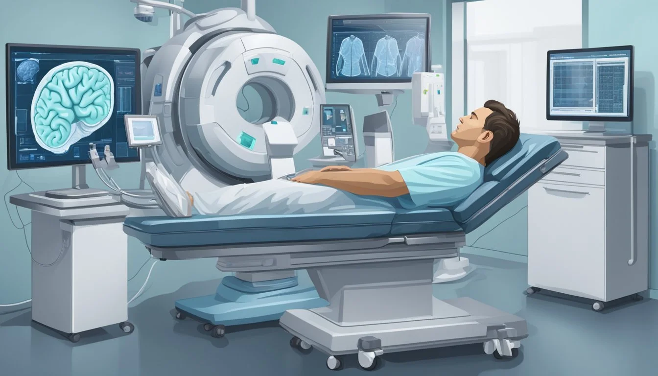
Neurological Scans to Diagnose Posttraumatic Stress Disorder
Brain imaging has revolutionized our understanding of posttraumatic stress disorder (PTSD). Over the past decade, researchers have utilized advanced neuroimaging techniques to examine the effects of traumatic stress on the brain. These studies have shed light on the neural circuits involved in PTSD and other stress-related disorders.
Neuroimaging studies have consistently implicated three key brain regions in PTSD: the amygdala, hippocampus, and medial prefrontal cortex. The amygdala, responsible for processing emotions and fear responses, often shows hyperactivity in individuals with PTSD. Conversely, the hippocampus, crucial for memory formation, and the medial prefrontal cortex, involved in emotional regulation, typically exhibit reduced activity and volume in PTSD patients.
Functional neuroimaging has provided valuable insights into the brain circuits mediating PTSD symptoms. These studies have revealed disrupted inhibitory signaling loops that normally help manage fear responses. By mapping these neural abnormalities, researchers aim to develop more targeted and effective treatments for PTSD, addressing the limitations of current psychotherapies and pharmacotherapies.
Fundamentals of PTSD
Posttraumatic Stress Disorder (PTSD) is a complex psychiatric condition that develops in response to traumatic events. It is characterized by specific symptom clusters and diagnostic criteria, with significant impacts on individuals and populations worldwide.
Defining PTSD and DSM-5 Criteria
PTSD is defined in the Diagnostic and Statistical Manual of Mental Disorders, Fifth Edition (DSM-5). The diagnosis requires exposure to actual or threatened death, serious injury, or sexual violence. This can occur through direct experience, witnessing the event, learning about it happening to a close friend or family member, or repeated exposure to aversive details.
Key diagnostic criteria include:
Intrusion symptoms (e.g., flashbacks, nightmares)
Avoidance of trauma-related stimuli
Negative alterations in cognition and mood
Changes in arousal and reactivity
Symptoms must persist for more than one month and cause significant distress or functional impairment.
Epidemiology of PTSD
PTSD affects a substantial portion of the global population. Lifetime prevalence rates vary:
General population: 6-8%
Combat veterans: 10-30%
Sexual assault survivors: 30-50%
Risk factors include:
Female gender
Lower socioeconomic status
Prior trauma exposure
Lack of social support
Certain occupations, such as military personnel, first responders, and healthcare workers, face increased risk due to higher trauma exposure.
Symptom Clusters and Clinical Presentation
PTSD symptoms are grouped into four distinct clusters:
Re-experiencing: Intrusive memories, nightmares, flashbacks
Avoidance: Efforts to avoid trauma-related thoughts, feelings, or reminders
Negative cognitions and mood: Persistent negative emotional state, diminished interest in activities
Hyperarousal: Irritable behavior, hypervigilance, exaggerated startle response
Clinical presentation can vary. Some individuals may experience predominantly avoidance symptoms, while others might struggle more with hyperarousal. Comorbid conditions like depression and substance use disorders are common.
PTSD can significantly impair social, occupational, and personal functioning. Early identification and appropriate treatment are crucial for improving outcomes and quality of life for those affected by this disorder.
PTSD and Comorbidities
Post-traumatic stress disorder (PTSD) frequently co-occurs with other mental and physical health conditions. These comorbidities can complicate diagnosis, treatment, and overall patient outcomes.
Common Comorbid Psychiatric Disorders
Major depressive disorder (MDD) is one of the most prevalent comorbidities in PTSD patients. Studies indicate that up to 50% of individuals with PTSD also meet criteria for MDD. This combination is associated with greater symptom severity and worse clinical outcomes than PTSD alone.
Anxiety disorders, particularly generalized anxiety disorder and panic disorder, are also common among PTSD sufferers. Substance use disorders affect a significant portion of PTSD patients, with many using alcohol or drugs to self-medicate symptoms.
Bipolar disorder and schizophrenia, while less frequent, can co-occur with PTSD. These complex comorbidities often require specialized treatment approaches.
Physical Health and PTSD
PTSD is linked to various physical health problems. Cardiovascular issues, including hypertension and heart disease, are more prevalent in PTSD patients. Chronic pain conditions, such as fibromyalgia and migraines, frequently co-exist with PTSD.
Gastrointestinal disorders, particularly irritable bowel syndrome, are common. Sleep disturbances, including insomnia and sleep apnea, affect many PTSD sufferers. These physical comorbidities can exacerbate PTSD symptoms and complicate treatment.
Addressing both mental and physical health concerns is crucial for comprehensive PTSD care. Integrated treatment approaches that target multiple comorbidities simultaneously often yield the best outcomes for patients.
Neurobiology of PTSD
Post-traumatic stress disorder (PTSD) involves complex neurobiological changes in the brain. These alterations affect key regions and networks involved in fear processing, emotion regulation, and memory formation.
Neural Correlates and Brain Regions
PTSD is associated with structural and functional changes in several brain areas. The amygdala, crucial for fear processing, often shows hyperactivity in PTSD patients. This heightened responsiveness contributes to exaggerated fear reactions and emotional dysregulation.
The prefrontal cortex (PFC), particularly the medial PFC, exhibits reduced activation in PTSD. This area is vital for emotion regulation and fear extinction. Its hypoactivity may explain difficulties in managing trauma-related memories and emotions.
Hippocampal damage is another hallmark of PTSD. Reduced hippocampal volume can impair contextual memory formation and retrieval, leading to overgeneralization of fear responses.
Neural Circuitry and Networks
PTSD affects multiple interconnected neural networks. The fear circuitry, involving the amygdala, hippocampus, and prefrontal cortex, shows altered connectivity and activation patterns.
The salience network, responsible for detecting and orienting to significant stimuli, often exhibits hyperactivity. This can result in heightened vigilance and exaggerated responses to potential threats.
The default mode network, involved in self-referential processing and autobiographical memory, may show disrupted functioning. This can contribute to difficulties in integrating traumatic memories into coherent narratives.
Neuroplasticity and Neurogenesis
PTSD can impact the brain's ability to adapt and form new neural connections. Chronic stress associated with PTSD may impair neuroplasticity, affecting learning and memory processes.
Hippocampal neurogenesis, the formation of new neurons in the hippocampus, can be suppressed in PTSD. This reduction may contribute to memory deficits and difficulties in contextualizing traumatic experiences.
Structural changes in the brain, such as reduced gray matter volume in key regions, can occur over time. These alterations may persist even after symptom improvement, highlighting the long-term neurobiological impact of PTSD.
Brain Imaging Techniques in PTSD
Brain imaging techniques have revolutionized our understanding of posttraumatic stress disorder (PTSD). These methods provide valuable insights into the structural and functional changes in the brain associated with PTSD.
Magnetic Resonance Imaging (MRI)
MRI plays a crucial role in structural neuroimaging for PTSD. It reveals focal gray matter atrophy in specific brain regions. These changes are often observed in the hippocampus, amygdala, and prefrontal cortex.
Structural MRI studies compare brain morphometry between PTSD patients and controls. They often include trauma-exposed non-PTSD individuals as a comparison group.
Diffusion Tensor Imaging (DTI), an MRI-based technique, measures white matter integrity. DTI studies in PTSD patients show altered fractional anisotropy in various brain regions.
Functional MRI (fMRI) and Its Applications
fMRI measures brain activity by detecting changes in blood oxygenation and flow. It has been instrumental in identifying altered neural activity and functional connectivity in PTSD.
Studies using fMRI have implicated several brain regions in PTSD pathophysiology:
Amygdala: Often shows hyperactivity
Hippocampus: Involved in memory processing
Medial prefrontal cortex: Plays a role in emotion regulation
fMRI tasks often involve trauma-related stimuli or emotional processing. These paradigms help elucidate the neural circuits mediating PTSD symptoms.
Positron Emission Tomography (PET)
PET imaging uses radioactive tracers to measure brain metabolism and neurotransmitter activity. It has been valuable in studying the neurochemical aspects of PTSD.
PET studies have shown:
Altered glucose metabolism in specific brain regions
Changes in neurotransmitter systems, particularly serotonin and norepinephrine
PET imaging can also assess treatment effects by measuring changes in brain activity before and after interventions.
Other Neuroimaging Modalities
Computed Tomography (CT) provides structural information but is less commonly used in PTSD research due to its lower resolution compared to MRI.
Single-Photon Emission Computed Tomography (SPECT) measures regional cerebral blood flow. It has been used to study PTSD but is less prevalent than PET or fMRI.
Magnetoencephalography (MEG) measures magnetic fields produced by neuronal activity. It offers high temporal resolution and has been used to study neural oscillations in PTSD.
Neuroimaging Studies in PTSD
Brain imaging techniques have revealed key neural structures involved in post-traumatic stress disorder (PTSD). These studies highlight alterations in the amygdala, hippocampus, and prefrontal cortex, shedding light on the neurobiology of PTSD symptoms.
Investigating the Amygdala and Fear Processing
Neuroimaging research has consistently shown increased amygdala activation in PTSD patients. This hyperactivity is associated with heightened fear responses and emotional reactivity. Functional MRI studies have revealed exaggerated amygdala responses to trauma-related stimuli and even neutral faces.
Resting-state functional connectivity analyses demonstrate altered amygdala connections with other brain regions in PTSD. These changes may contribute to difficulties in fear extinction, a process crucial for recovery from trauma.
PET and SPECT studies have also found increased blood flow and metabolism in the amygdala of PTSD patients, further supporting its role in the disorder's pathophysiology.
The Role of the Hippocampus and Memory
Structural neuroimaging studies have consistently reported reduced hippocampal volume in PTSD patients. This reduction may explain memory deficits and altered contextual processing observed in the disorder.
Functional imaging has shown decreased hippocampal activation during memory tasks in PTSD. This finding aligns with difficulties in forming and retrieving coherent memories of traumatic events.
Longitudinal studies suggest that smaller hippocampal volume may be both a risk factor for developing PTSD and a consequence of chronic stress exposure. This bidirectional relationship highlights the complexity of PTSD neurobiology.
Prefrontal Cortex and Emotion Regulation
Neuroimaging studies have revealed decreased activation in the medial prefrontal cortex (mPFC) and anterior cingulate cortex (ACC) in PTSD patients. These regions are crucial for emotion regulation and fear extinction.
Reduced prefrontal activation is often accompanied by increased amygdala activity, suggesting a disruption in the normal inhibitory control of emotional responses. This imbalance may contribute to the persistent re-experiencing of traumatic memories.
Functional connectivity between the mPFC and amygdala is often altered in PTSD, potentially explaining difficulties in modulating fear responses. Treatment studies have shown that successful interventions can normalize this connectivity, highlighting potential neural targets for PTSD therapies.
Clinical Implications and Treatment Correlations
Brain imaging studies have revealed valuable insights into PTSD, guiding treatment approaches and improving outcome predictions. Neuroimaging findings are increasingly shaping personalized interventions and informing the development of novel therapies.
Tailoring Treatment Based on Neuroimaging
Brain imaging allows clinicians to customize PTSD treatments based on individual neural patterns. Patients with hyperactive amygdala responses may benefit from exposure-based therapies to reduce fear reactivity. Those showing prefrontal cortex deficits often respond well to cognitive restructuring techniques.
Neuroimaging can identify specific brain circuits affected by trauma, enabling targeted interventions. For example, transcranial magnetic stimulation (TMS) can be precisely aimed at underactive prefrontal regions to enhance emotional regulation.
Some clinics now use functional MRI to track treatment progress, adjusting therapy plans based on observed brain changes. This approach helps optimize treatment duration and intensity for each patient.
Predicting Treatment Outcomes
Brain imaging markers show promise in forecasting PTSD treatment responses. Pre-treatment amygdala reactivity often predicts outcomes for exposure therapy. Lower hippocampal volume is associated with poorer prognosis across various interventions.
Neuroimaging can identify patients likely to benefit from specific treatments. Those with heightened anterior cingulate activity tend to respond better to cognitive-behavioral therapy. Conversely, reduced anterior cingulate function may indicate a need for medication-based approaches.
Predictive models combining brain imaging data with clinical assessments are being developed. These tools aim to guide treatment selection, potentially improving success rates and reducing the trial-and-error process often experienced by patients.
Advances in Pharmacotherapy and Psychotherapy
Brain imaging studies are driving innovations in PTSD medications. New drugs targeting the reconsolidation of traumatic memories show promise in reducing amygdala hyperactivity. Ketamine's rapid antidepressant effects have been linked to increased prefrontal connectivity, spurring research into similar compounds.
Psychotherapy techniques are evolving based on neuroimaging insights. Virtual reality exposure therapy now incorporates real-time fMRI feedback to optimize fear extinction. Mindfulness-based interventions are being refined to enhance activation of emotion regulation circuits.
Neurofeedback training, guided by EEG or fMRI data, allows patients to directly modulate brain activity linked to PTSD symptoms. This approach shows potential in reducing hyperarousal and improving emotional control.
Emerging Techniques and Future Directions
Brain imaging for PTSD is advancing rapidly with new technologies and approaches. These innovations aim to improve diagnosis, treatment, and understanding of the disorder's underlying neurobiology.
Machine Learning in PTSD Diagnosis
Machine learning algorithms are revolutionizing PTSD diagnosis through neuroimaging data analysis. These sophisticated tools can identify subtle patterns in brain structure and function that may be imperceptible to human observers.
Researchers are developing models that combine multiple imaging modalities, such as fMRI and DTI, to enhance diagnostic accuracy. These algorithms can differentiate PTSD from other psychiatric disorders with increasing precision.
Recent studies have shown promising results in using machine learning to predict treatment outcomes. This approach could help clinicians tailor interventions to individual patients based on their unique neural profiles.
Innovative Brain Stimulation Approaches
Novel brain stimulation techniques are emerging as potential treatments for PTSD. Transcranial magnetic stimulation (TMS) and transcranial direct current stimulation (tDCS) show promise in modulating neural activity in key brain regions.
These non-invasive methods target specific neural circuits implicated in PTSD, such as the amygdala and prefrontal cortex. By altering activity in these areas, researchers aim to reduce symptoms and improve emotional regulation.
Closed-loop neurofeedback systems are another exciting development. These devices monitor brain activity in real-time and deliver targeted stimulation when abnormal patterns are detected, potentially interrupting traumatic memories or anxiety responses.

